


Effect of Prolonged Fasting on Uric Acid Metabolism and Its Regulation Mechanism
-
摘要: 为研究长期禁食过程中大鼠尿酸代谢的变化及其潜在的调控机制,以Sprague-Dawley(SD)大鼠为动物模型,通过病理组织切片、生化检测、荧光定量PCR(qRT-PCR)以及蛋白免疫印迹(Western blotting)等方法分析不同禁食时间(1,2,3,5,7天)大鼠尿酸水平及其代谢相关基因和蛋白的表达变化。结果表明,长期禁食未对大鼠肾脏组织产生明显的损伤,引起了血尿酸水平上升、尿尿酸水平波动性变化和血液尿酸酶活性升高;随着禁食时间的延长,主要尿酸转运蛋白的mRNA和蛋白表达水平逐渐上调。长期禁食过程中大鼠尿酸代谢变化可能与尿酸转运蛋白及尿酸酶活性有关。Abstract: To investigate the changes of uric acid metabolism in rats during prolonged fasting and its potential regulatory mechanism. Sprague-Dawley (SD) rats were used as animal models. The changes of uric acid level and metabolism-related gene and protein expression in rats during different fasting periods (1, 2, 3, 5, and 7 days) were analyzed by pathological tissue sections, biochemical tests, quantitative PCR (qRT-PCR), and western blotting. The results showed that prolonged fasting did not cause significant damage to kidney tissue, but remarkedly increased the blood uric acid level and blood uricase activity. It also resulted in fluctuating changes in urine uric acid level. The mRNA and protein expression levels of main uric acid transporters were increased gradually with the fasting duration. The elevation of serum uric acid caused by prolonged fasting is related to uric acid transporter and uricase activity.
-
Key words:
- Prolonged fasting /
- Uric acid metabolism /
- Regulatory mechanism /
- Hypometabolism
-

图 1 不同禁食时间各组大鼠肾病理形态学变化(n=3)。(a)正常对照组肾脏,(b)禁食1天肾脏,(c)禁食2天肾脏,(d)禁食3天肾脏,(e)禁食5天肾脏,(f)禁食7天肾脏。红色箭头表示肾小球,蓝色箭头表示肾小管上皮细胞,黑色箭头表示上皮细胞脱落及炎症细胞浸润
Figure 1. Pathological changes of the kidneys during different fasting duration in rats (n=3 in each group). (a) Normal control group, (b) fasting for 1 day, (c) fasting for 2 days, (d) fasting for 3 days, (e) fasting for 5 days, (f) fasting for 7 days. Red arrow: glomerulus. Blue arrow: tubular epithelial cells. Black arrow: epithelial cell exfoliation and inflammatory cell infiltration

图 2 不同禁食时间各组大鼠肾脏组织病理形态学变化检测评分(n=3)
Figure 2. Histological scores in kidneys during different fasting duration in rats (n=3 in each group)

图 3 不同禁食时间大鼠血肌酐(a)和尿素(b)变化。n=6,**P<0.01,与 0 d(对照组)比较
Figure 3. Changes of blood creatinine (a) and urea (b) during different fasting duration in rats. n=6, **P<0.01, compared with 0 d (control group)

图 4 不同禁食时间大鼠血尿酸(a)和尿尿酸(b)变化。n=6,*P<0.05,**P<0.01,与 0 d(对照组)比较
Figure 4. Changes of blood uric acid (a) and urine uric acid (b) during different fasting duration in rats. n=6, *P<0.05, **P<0.01, compared with 0 d (control group)

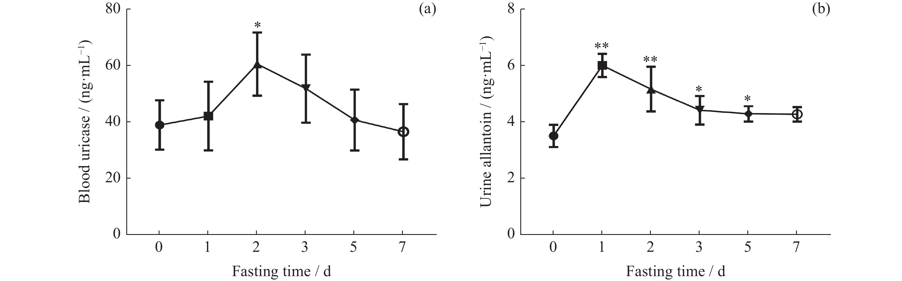
图 5 不同禁食时间大鼠血液尿酸酶(a)和尿液尿囊素(b)水平变化。n=6,*P<0.05,**P<0.01,与 0 d(对照组)比较
Figure 5. Changes of blood uricase (a) and urine allantoin (b) during different fasting duration in rats. n=6, *P<0.05, **P<0.01, compared with 0 d (control group)

图 6 不同禁食时间大鼠尿酸转运蛋白mRNA表达水平变化。n=6,*P<0.05,**P<0.01,与 0 d(对照组)比较。(a) GLUT9,(b) URAT1,(c) OAT1,(d) Mrp4,(e) UAT,(f) NPT1
Figure 6. mRNA changes of urate transporter during different fasting duration in rats. n=6, *P<0.05, **P<0.01, compared with 0 d (control group). (a) GLUT9, (b) URAT1, (c) OAT1, (d) Mrp4, (e) UAT, (f) NPT1

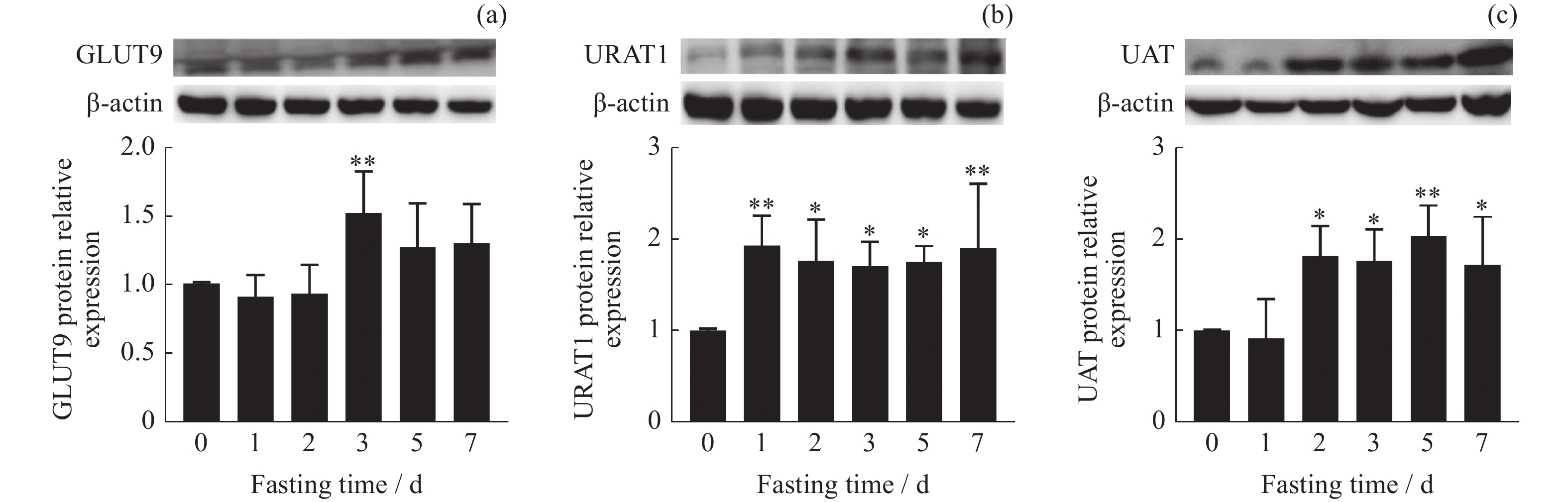
图 7 不同禁食时间大鼠主要尿酸转运蛋白(GLUT9,URAT1,UAT)的蛋白表达水平变化。n=6,*P<0.05,**P<0.01,与 0 d(对照组)比较。(a)GLUT9蛋白相对表达量,(b)URAT1蛋白相对表达量,(c)UAT蛋白相对表达量
Figure 7. Protein changes of major urate transporter (GLUT9, URAT1, UAT) during different fasting duration in rats. n=6, *P<0.05, **P<0.01, compared with 0 d (control group). (a) The protein relative expression of GLUT9 protein, (b) the protein relative expression of URAT1 protein, (c) the protein relative expression of UAT protein

图 8 不同禁食时间大鼠血尿酸与主要尿酸转运蛋白mRNA和蛋白相对表达量之间相关性分析。血尿酸与尿酸转运蛋白GLUT9 (a),URAT1(b),UAT(c) mRNA相对表达量,以及GLUT9(d),URAT1(e),UAT(f)蛋白相对表达量之间相关性分析
Figure 8. Correlation analysis between blood uric acid and mRNA and protein relative expression of main uric acid transporter during different fasting duration in rats. The correlation analysis between blood uric acid and the mRNA expression and protein expression of (a)(d) GLUT9, (b)(e) URAT1, (c)(f) UAT
-
[1] 戴钟铨, 李莹辉, 杨超, 等. 面向未来载人星际航行的空间低代谢调节技术[J]. 载人航天, 2021, 27(3): 269-275 doi: 10.16329/j.cnki.zrht.2021.03.001DAI Zhongquan, LI Yinghui, YANG Chao, et al. Space hypometabolic regulation technology for future manned interplanetary flight[J]. Manned Spaceflight, 2021, 27(3): 269-275 doi: 10.16329/j.cnki.zrht.2021.03.001 [2] PAK H H, HAWS S A, GREEN C L, et al. Fasting drives the metabolic, molecular and geroprotective effects of a calorie-restricted diet in mice[J]. Nature Metabolism, 2021, 3(10): 1327-1341 doi: 10.1038/s42255-021-00466-9 [3] DE CABO R, MATTSON M P. Effects of intermittent fasting on health, aging, and disease[J]. The New England Journal of Medicine, 2019, 381(26): 2541-2551 doi: 10.1056/NEJMra1905136 [4] ANTON S D, MOEHL K, DONAHOO W T, et al. Flipping the metabolic switch: understanding and applying the health benefits of fasting[J]. Obesity, 2018, 26(2): 254-268 doi: 10.1002/oby.22065 [5] MATTSON M P, LONGO V D, HARVIE M. Impact of intermittent fasting on health and disease processes[J]. Ageing Research Reviews, 2017, 39: 46-58 doi: 10.1016/j.arr.2016.10.005 [6] MANDAL A K, MOUNT D B. The molecular physiology of uric acid homeostasis[J]. Annual Review of Physiology, 2015, 77: 323-345 doi: 10.1146/annurev-physiol-021113-170343 [7] DE TOLEDO F W, GRUNDLER F, BERGOUIGNAN A, et al. Safety, health improvement and well-being during a 4 to 21-day fasting period in an observational study including 1422 subjects[J]. PLoS One, 2019, 14(1): e0209353 doi: 10.1371/journal.pone.0209353 [8] MOJTO V, GVOZDJAKOVA A, KUCHARSKA J, et al. Effects of complete water fasting and regeneration diet on kidney function, oxidative stress and antioxidants[J]. Bratislava Medical Journal, 2018, 119(2): 107-111 doi: 10.4149/BLL_2018_020 [9] CAMELO L, DE SOUZA MARINHO T, ÁGUILA M B, et al. Intermittent fasting exerts beneficial metabolic effects on blood pressure and cardiac structure by modulating local renin-angiotensin system in the heart of mice fed high-fat or high-fructose diets[J]. Nutrition Research, 2019, 63: 51-62 doi: 10.1016/j.nutres.2018.12.005 [10] LI L J, ZHANG Y P, ZENG C C. Update on the epidemiology, genetics, and therapeutic options of hyperuricemia[J]. American Journal of Translational Research, 2020, 12(7): 3167-3181 [11] SPITSIN S V, SCOTT G S, MIKHEEVA T, et al. Comparison of uric acid and ascorbic acid in protection against EAE[J]. Free Radical Biology and Medicine, 2002, 33(10): 1363-1371 doi: 10.1016/s0891-5849(02)01048-1 [12] AMES B N, CATHCART R, SCHWIERS E, et al. Uric acid provides an antioxidant defense in humans against oxidant- and radical-caused aging and cancer: a hypothesis[J]. Proceedings of the National Academy of Sciences of the United States of America, 1981, 78(11): 6858-6862 doi: 10.1073/pnas.78.11.6858 [13] SHI Y, EVANS J E, ROCK K L. Molecular identification of a danger signal that alerts the immune system to dying cells[J]. Nature, 2003, 425(6957): 516-521 doi: 10.1038/nature01991 [14] WANG Z, CUI T, CI X Y, et al. The effect of polymorphism of uric acid transporters on uric acid transport[J]. Journal of Nephrology, 2019, 32(2): 177-187 doi: 10.1007/s40620-018-0546-7 [15] SATO Y, FEIG D I, STACK A G, et al. The case for uric acid-lowering treatment in patients with hyperuricaemia and CKD[J]. Nature Reviews Nephrology, 2019, 15(12): 767-775 doi: 10.1038/s41581-019-0174-z [16] XU L Q, SHI Y F, ZHUANG S G, et al. Recent advances on uric acid transporters[J]. Oncotarget, 2017, 8(59): 100852-100862 doi: 10.18632/oncotarget.20135 [17] GABEL K, KROEGER C M, TREPANOWSKI J F, et al. Differential effects of alternate-day fasting versus daily calorie restriction on insulin resistance[J]. Obesity, 2019, 27(9): 1443-1450 doi: 10.1002/oby.22564 [18] PHILLIPS M C L. Fasting as a therapy in neurological disease[J]. Nutrients, 2019, 11(10): 2501 doi: 10.3390/nu11102501 [19] LI C Y, OSTERMANN T, HARDT M, et al. Metabolic and psychological response to 7-day fasting in obese patients with and without metabolic syndrome[J]. Forschende Komplementarmedizin, 2013, 20(6): 413-420 doi: 10.1159/000353672 [20] XIONG Q, LIU J, XU Y C. Effects of uric acid on diabetes mellitus and its chronic complications[J]. International Journal of Endocrinology, 2019, 2019: 9691345 doi: 10.1155/2019/9691345 [21] CORTESE F, GIORDANO P, SCICCHITANO P, et al. Uric acid: from a biological advantage to a potential danger. A focus on cardiovascular effects[J]. Vascular Pharmacology, 2019, 120: 106565 doi: 10.1016/j.vph.2019.106565 [22] EL DIN U A A S, SALEM M M, ABDULAZIM D O. Uric acid in the pathogenesis of metabolic, renal, and cardiovascular diseases: a review[J]. Journal of Advanced Research, 2017, 8(5): 537-548 doi: 10.1016/j.jare.2016.11.004 [23] VEGA J, HUIDOBRO E J P. Effects of creatine supplementation on renal function[J]. Revista Médica de Chile, 2019, 147(5): 628-633 doi: 10.4067/S0034-98872019000500628 [24] ENSMINGER D C, SALVADOR-PASCUAL A, ARANGO B G, et al. Fasting ameliorates oxidative stress: a review of physiological strategies across life history events in wild vertebrates[J]. Comparative Biochemistry and Physiology Part A: Molecular & Integrative Physiology, 2021, 256: 110929 doi: 10.1016/j.cbpa.2021.110929 [25] BRAGA T T, FORESTO-NETO O, CAMARA N O S. The role of uric acid in inflammasome-mediated kidney injury[J]. Current Opinion in Nephrology and Hypertension, 2020, 29(4): 423-431 doi: 10.1097/MNH.0000000000000619 [26] RICHETTE P, BARDIN T. Gout[J]. The Lancet, 2010, 375(9711): 318-328 doi: 10.1016/S0140-6736(09)60883-7 [27] LIU X D. SLC family transporters[J]. Advances in Experimental Medicine and Biology, 2019, 1141: 101-202 doi: 10.1007/978-981-13-7647-4_3 [28] SHIBASAKI K, KIMURA M, IKARASHI R, et al. Uric acid concentration in saliva and its changes with the patients receiving treatment for hyperuricemia[J]. Metabolomics, 2012, 8(3): 484-491 doi: 10.1007/s11306-011-0334-z [29] BU P L, LE Y, ZHANG Y, et al. Hormonal and chemical regulation of the Glut9 transporter in mice[J]. Journal of Pharmacology and Experimental Therapeutics, 2017, 360(1): 206-214 doi: 10.1124/jpet.116.237040 [30] UEMURA N, MURAKAMI R, CHIBA Y, et al. Immunoreactivity of urate transporters, GLUT9 and URAT1, is located in epithelial cells of the choroid plexus of human brains[J]. Neuroscience Letters, 2017, 659: 99-103 doi: 10.1016/j.neulet.2017.09.001 [31] BOBULESCU I A, MOE O W. Renal transport of uric acid: evolving concepts and uncertainties[J]. Advances in Chronic Kidney Disease, 2012, 19(6): 358-371 doi: 10.1053/j.ackd.2012.07.009 -
-

 下载:
下载:


计量
- 文章访问数: 2341
- HTML全文浏览量: 995
- PDF下载量: 53
-
被引次数:
0(来源:Crossref)
0(来源:其他)

